
Digital Pathology
Application
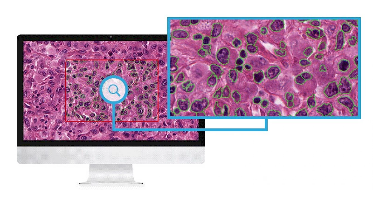
The digital pathology system is based on four key components:
- Capturing images (of slides) using digital scanners: Glass slides are represented in the digital form by using whole slide scanning, along with proper tools in an indicative manner. High-resolution scanning is performed, along with adequate color depth, so that images can be reproduced with the necessary magnifications required for diagnostic and research applications.
- Storing and archiving the images: The strategy of storing virtual slides is mainly dependent on the intended use. certain instances, off-site storage may be required for remote consultation, using different options, optical/tape-based, cloud storage, or a combination approach.
Portuguese and European Collaborative Programs and Projects are running
- Editing and modifying captured images: Digital slides provide the opportunity to modify images according to the pathologist’s requirement, such as magnification, zoom-in/out, etc.
- Viewing and sharing images: Digital slides can be easily shared with teacher-students, doctors, research institutes, etc.